
History and discussion
Presentation
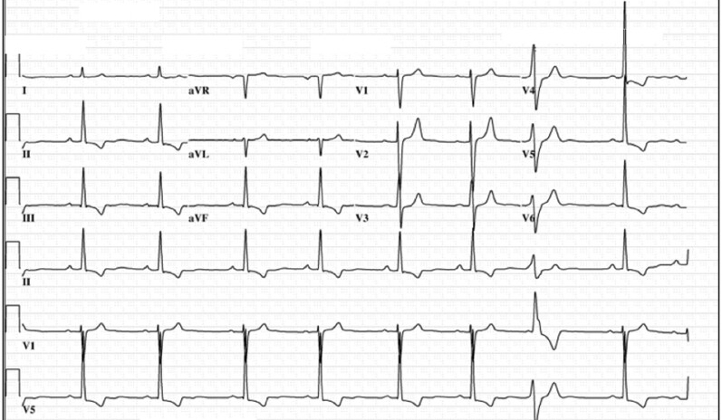
38 yrs old lady had 2 episodes of syncope- one while walking and other while sitting. Her ECG showed sinus rhythm with T wave inversions in inferior leads with normal PR interval and QRSD of 100 msec and normal Qtc. Echo showed mild degree of mitral valve prolapse but with normal bi ventricular function. ELR 3 days was uneventful except for low burden of ventricular ectopy with no symptoms during ectopy. HUTT was negative. How would you proceed to investigate this lady.
Presentation
- 38 year old lady
- Two episodes of syncope
- T inversion in inferior leads
- Mild MVP, Normal LV function
- Three day ELR - Low burden of asymptomatic ventricular ectopy
- HUTT negative
Discussion - Review of previous investigations
- Detailed history
- ECG - what else to look for ?
- Echo review - what to look for ?
- ELR review - what to look for ?
Discussion - Additional investigations
- Implantable loop recorder ?
- Is CMR required ?
- Is EP study indicated ?
Discussion - Management
- How will I discuss prognosis ?
- What is the risk of sudden death ?
- Is ICD indicated ?
- Is ablation to be considered ?
Review
History
- Syncope during exertion
- Prodrome
- Associated or preceding palpitations
- Injury
ECG
Echo review
- Confirm diagnosis of MVP
- Characterise MVP
- Assess for MAD
- Quantification of MR
- LV function and dimensions
Diagnosis of MVP
- Superior displacement of the mitral leaflet(s)
- >2 mm above annular plane during systole
- Parasternal long axis view
Characterise MVP
- Bileaflet
- Myxomatous degeneration - maximal leaflet thickness of at least 5 mm
Annular mid-systolic spike (Pickelhaube sign) > 16 cm/s
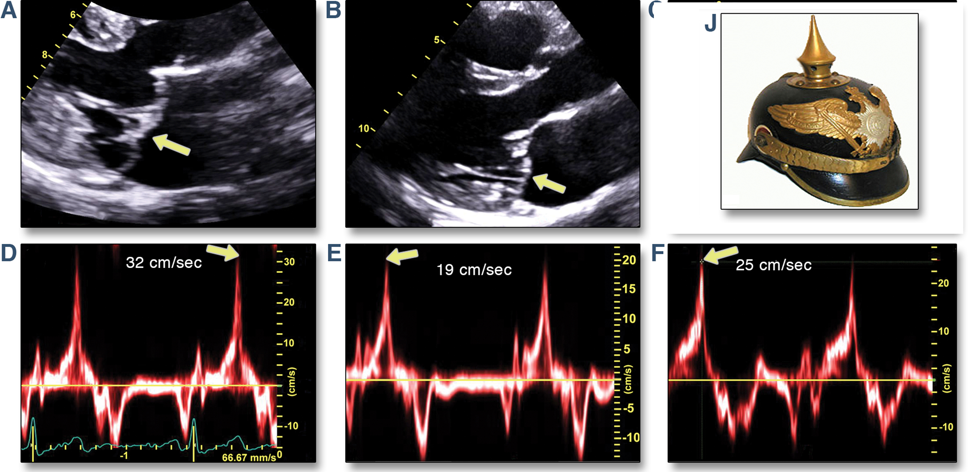
Muthukumar L., Rahman F., Jan M.F. et al: The Pickelhaube sign: novel echocardiographic risk marker for malignant mitral valve prolapse syndrome. J Am Coll Cardiol Img 2017; 10: 1078.
Mitral annular dysjunction
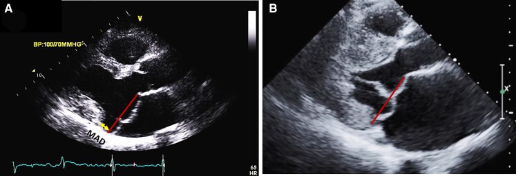
- Systolic separation between the ventricular myocardium and the mitral annulus supporting the posterior mitral leaflet
- Loss of mechanical annular function
Assess for MAD
- Parasternal long axis at end-systole
- Distance from LA wall-MV junction to top of LV myocardium
- Highest risk for MAD at posterior wall
- Systolic curling motion
Mitral annular dysjunction
Long term ECG recording
- Confirm beat templates
- Monomorphic vs polymorphic PVCs
- Couplets / NSVT
Additional investigations
ILR ?
- Longer ECG recording may uncover more arrhythmias
- May provide symptom - rhythm correlation
- Will advise if not implanting ICD
CMR
- Confirms findings of echocardiography
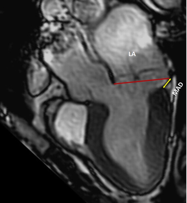
- Assessment of MAD (six long axis cine sequences)
- Fibrosis assessment
MVP assessment by CMR
Fibrosis
- LGE - Especially involving annulus / papillary muscles
- Diffuse LV fibrosis with T1 mapping
EP study
- No robust data
- Not usually useful
- Induction of monomorphic VT could have more value
Management
Prognosis
- Common, generally benign in absence of significant MR
- Small subset have higher risk of VA and SCD
- Related to arrhythmias
Early recognition of sudden death risk
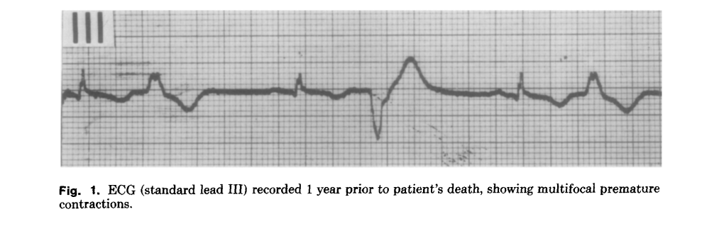
Pocock WA, Bosman CK, Chesler E, Barlow JB, Edwards JE. Sudden death in primary mitral valve prolapse. Am Heart J. 1984 Feb;107(2):378-82. doi: 10.1016/0002-8703(84)90389-2. PMID: 6695671.
Prospective data on outcomes in MVP
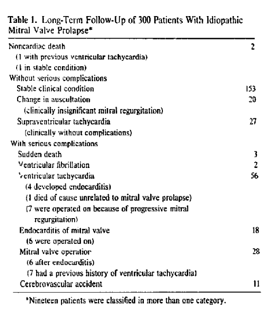
Düren DR, Becker AE, Dunning AJ. Long-term follow-up of idiopathic mitral valve prolapse in 300 patients: a prospective study. J Am Coll Cardiol. 1988 Jan;11(1):42-7. doi: 10.1016/0735-1097(88)90164-7.
Prospective study - Arrhythmias and mortality
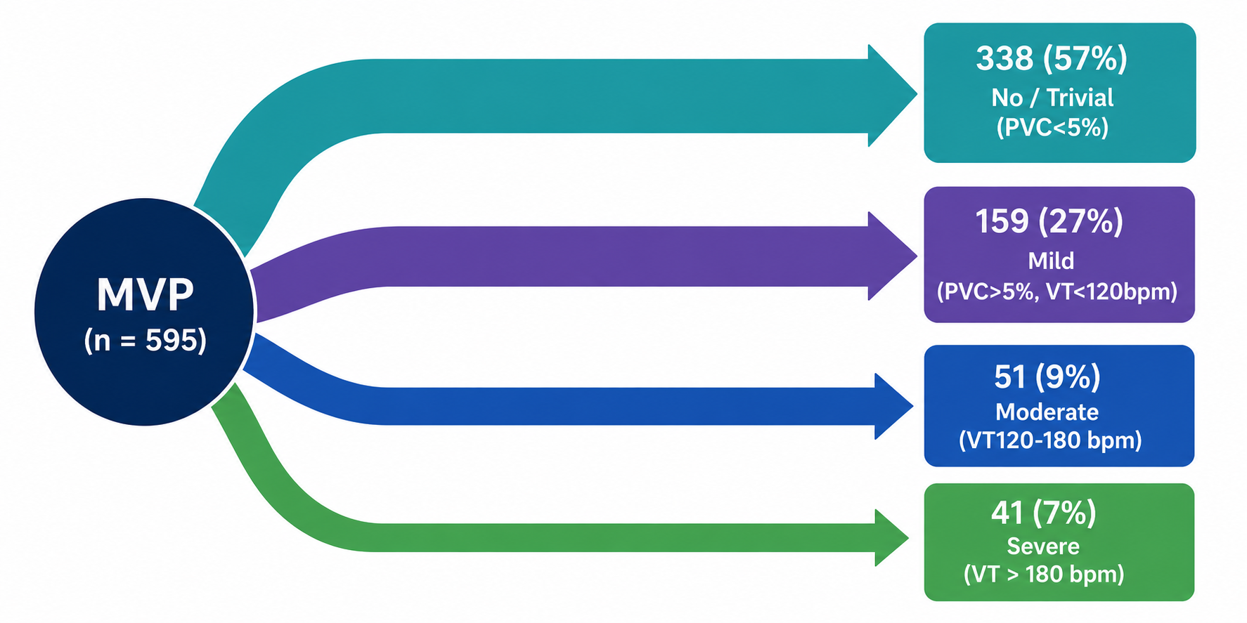
Essayagh B, Sabbag A, Antoine C, Benfari G, Yang LT, Maalouf J, Asirvatham S, Michelena H, Enriquez-Sarano M. Presentation and Outcome of Arrhythmic Mitral Valve Prolapse. J Am Coll Cardiol. 2020 Aug 11;76(6):637-649. doi: 10.1016/j.jacc.2020.06.029. PMID: 32762897.
Mortality at 4 years
- No / Trivial - 6%
- Mild to Moderate - 9%
- Severe - 14%
Drug therapy
- Beta blockers may reduce PVCs
- AAD if PVC related LV dysfunction - Flecainide, Amiodarone or Propafenone
- Quinidine for Purkinje origin short coupled PVCs
Ablation
- PVC induced VF
- PVC induced cardiomyopathy
- Sustained VT with ICD therapies
PVC / VT origin
- Papillary muscles
- Outflow tract
- Purkinje origin PVC
Miller MA, Dukkipati SR, Turagam M, Liao SL, Adams DH, Reddy VY. Arrhythmic Mitral Valve Prolapse: JACC Review Topic of the Week. J Am Coll Cardiol. 2018 Dec 11;72(23 Pt A):2904-2914. doi: 10.1016/j.jacc.2018.09.048. PMID: 30522653.
Purkinje ectopy
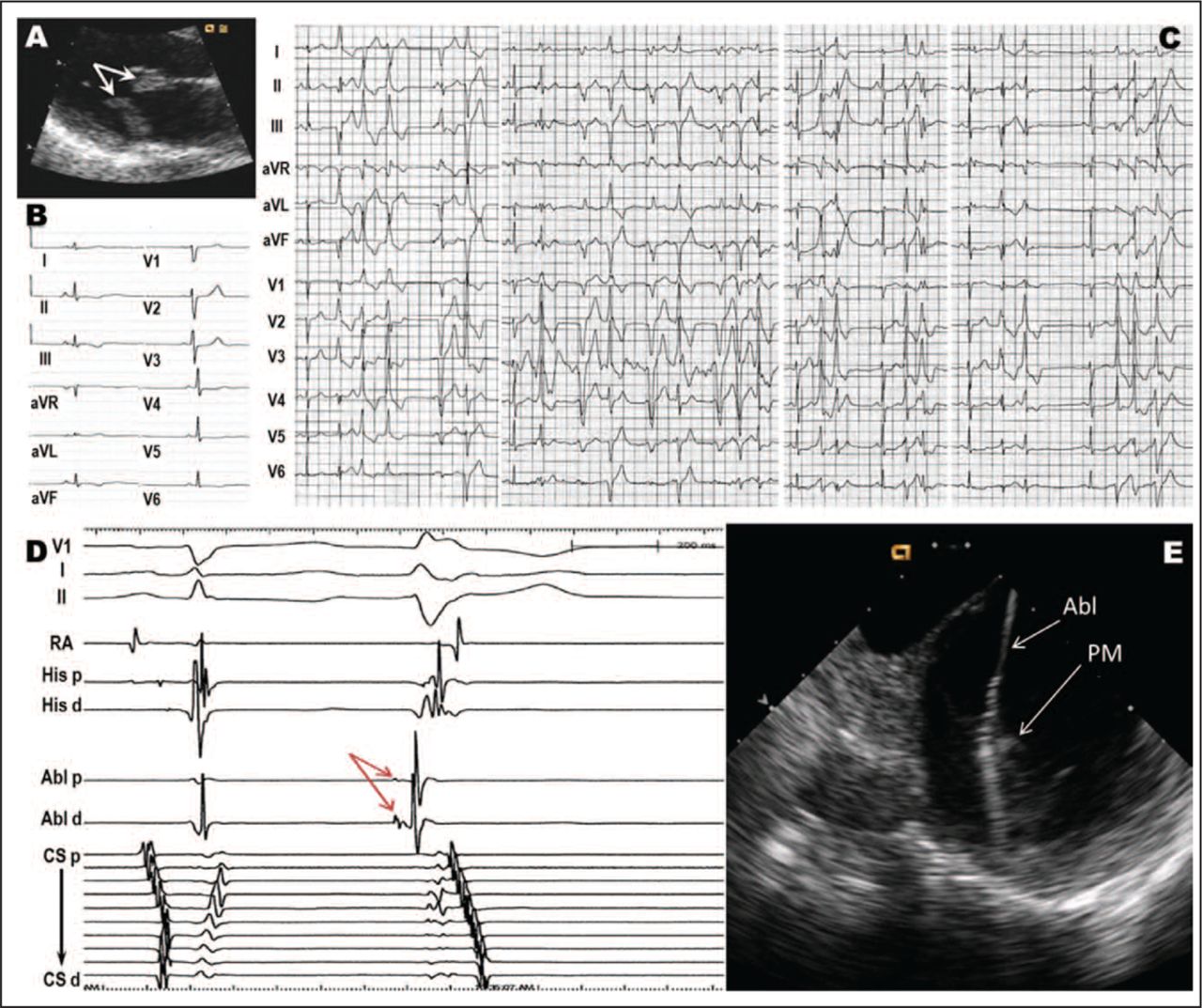
Syed FF, Ackerman MJ, McLeod CJ, Kapa S, Mulpuru SK, Sriram CS, Cannon BC, Asirvatham SJ, Noseworthy PA. Sites of Successful Ventricular Fibrillation Ablation in Bileaflet Mitral Valve Prolapse Syndrome. Circ Arrhythm Electrophysiol. 2016 May;9(5):e004005. doi: 10.1161/CIRCEP.116.004005.
Risk stratification
Causes of SCD
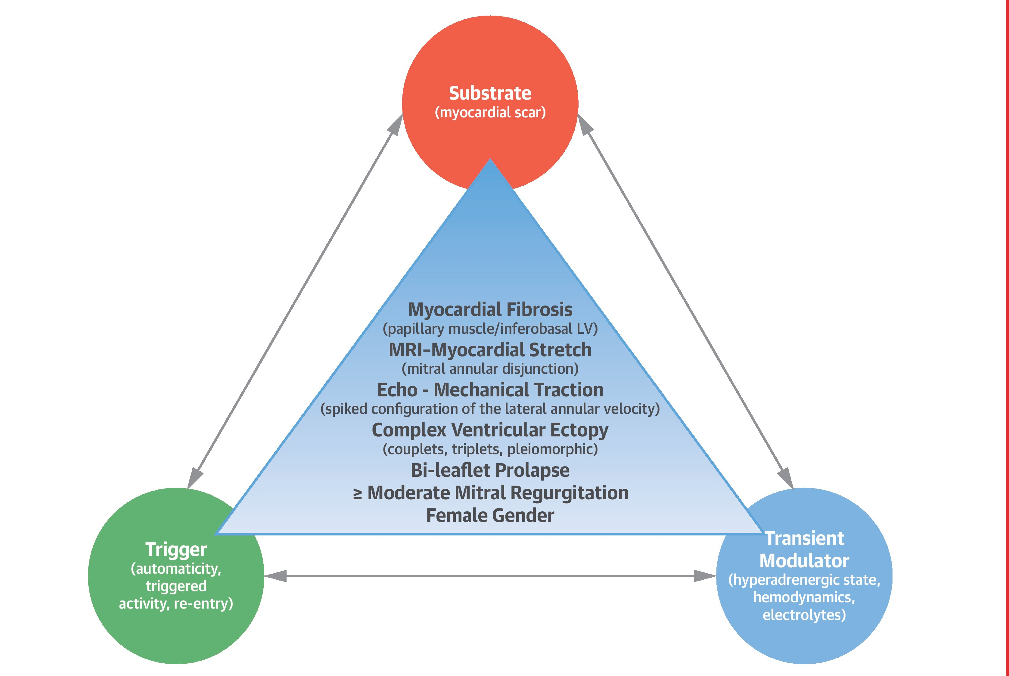
Miller MA, Dukkipati SR, Turagam M, Liao SL, Adams DH, Reddy VY. Arrhythmic Mitral Valve Prolapse: JACC Review Topic of the Week. J Am Coll Cardiol. 2018 Dec 11;72(23 Pt A):2904-2914. doi: 10.1016/j.jacc.2018.09.048.
MVP in OHCA
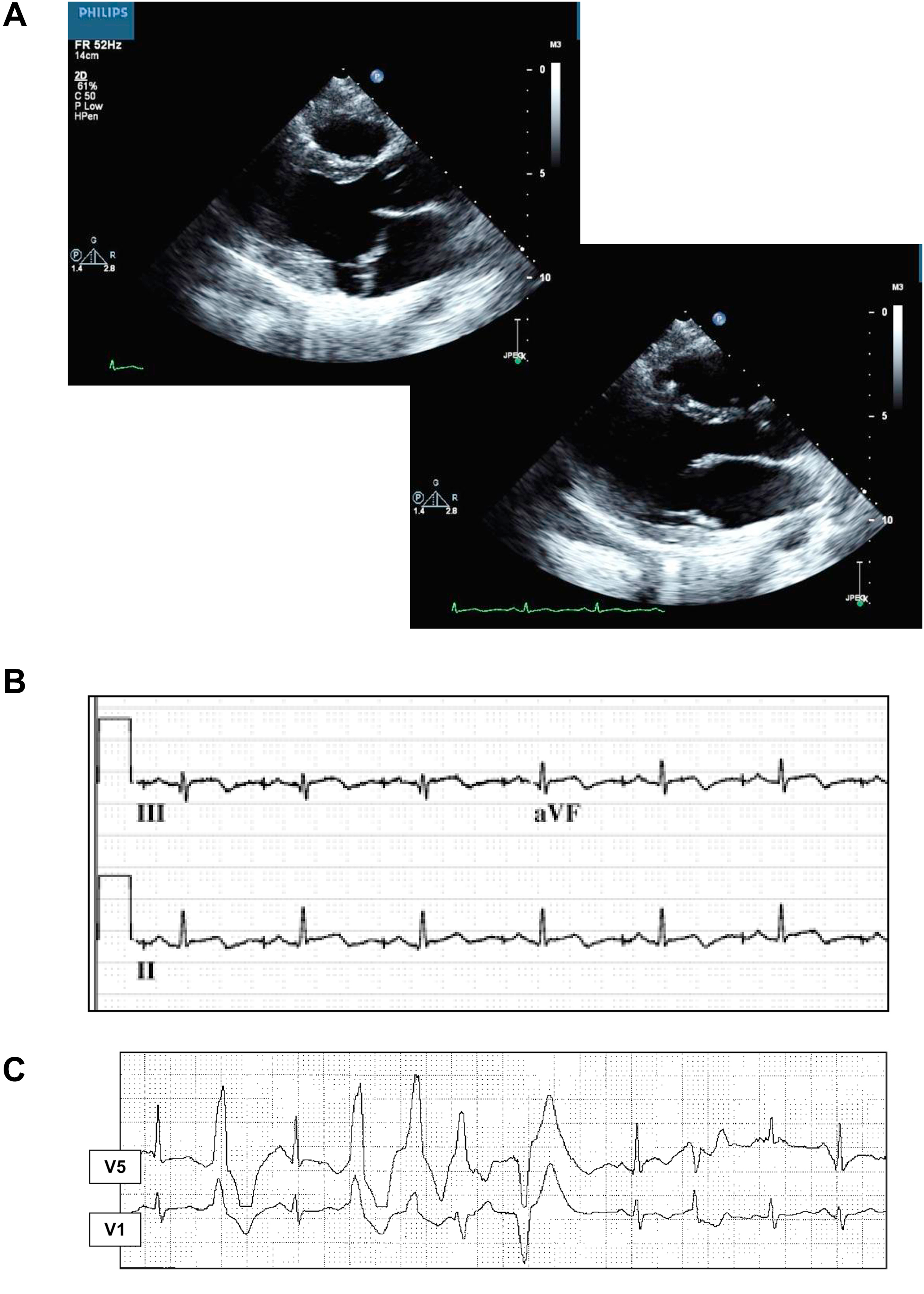
- More Bileaflet MVP
- Biphasic / inverted T waves
- More ventricular bigemini and VT in Holter
- PVCs from outflow alternating with papillary / fascicular
ICD indications
- Secondary prevention - Documented VA / Cardiac arrest
- Primary prevention - LVEF < 35%
- Unexplained syncope and high risk VAs
- One high risk feature and 2 or more phenotypic features
- High risk - Unexplained syncope, NSVT, sustained VT
- Phenotypic features - Polymorphic PVCs, MAD, LGE …
EHRA expert consensus statement on arrhythmic mitral valve prolapse and mitral annular disjunction complex. Europace. 2022 Dec 9;24(12):1981-2003. doi: 10.1093/europace/euac125.
Summary
- Careful history for syncope cause
- Echo review
- MRI heart
- ICD if other high risk features
- ILR otherwise
Summary
- MVP in general is common and benign
- Subset at risk of arrhythmias and sudden death
- Unexplained syncope is a high risk marker
- Prompts aggressive evaluation for arrhythmias and ICD if indicated